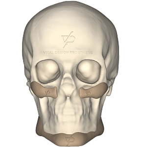
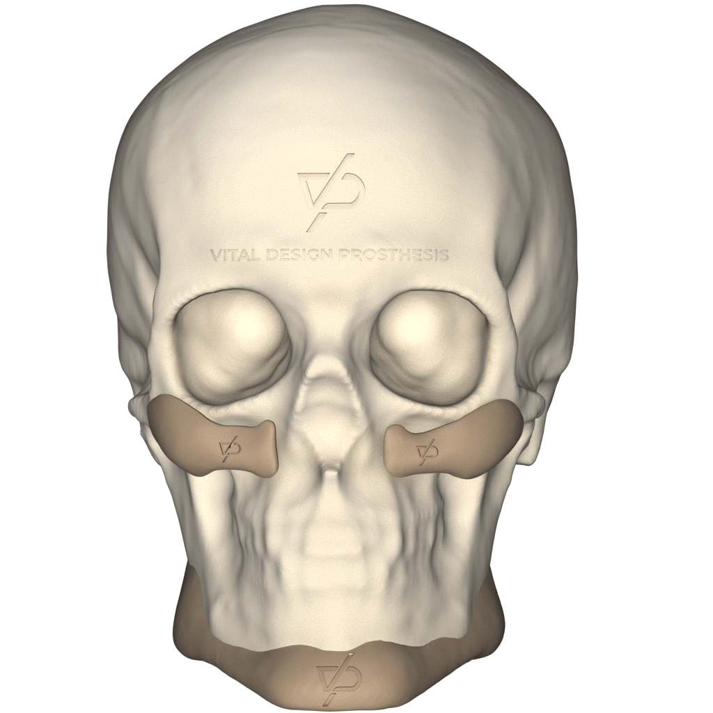
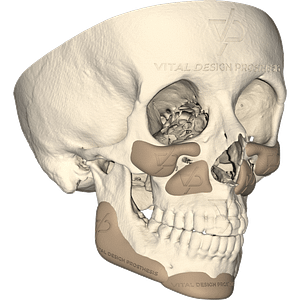
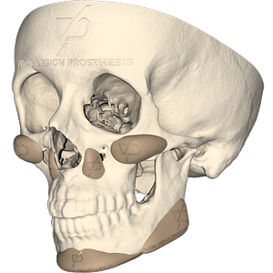
The personalized facial augmentation implants manufactured in VDP are made from the DICOM images of the patient’s Computed Tomography.
They are used to provide a greater contour and facial harmony, correcting facial asymmetries, rejuvenating and providing a balance and increase to different areas of the face, such as: chin, mandibular angle, malar, submalar and the infraorbital and paranasal areas.
PEEK is the main material used for the production of the implants and have a long clinical history of more than 20 years of use.
Benefits
Patient
-Improves quality of life
-Natural aesthetic appearance
-Bone support
-Decreases the time of rehabilitation
-General cost reduction
Surgeons
-Perfect fit of the implant
-Reduction in surgical time
-Faster and easier surgery
-Reduces the risk of infection and complications
Hospital
-Shorter hospital stays
-Lower cost of treatment
Distinctive Features of PEEK
PEEK is a biomaterial used since the 2000s, with a successful history in the correction of craniomaxillofacial defects.
It presents an excellent biocompatibility with adjacent tissues, decreasing inflammatory reactions.
Due to their great malleability, they can be modified in situ with drill bits.
It is radiopaque, which allows its visibility in postoperative scans.
Indications of use
-Improve facial harmony
-Improve facial contour
-Malar hypoplasia
-Facial asymmetry
-Assistant in orthognathic surgery
-Congenital diseases
Types of Facial Augmentation Implant
-Chin
-Mandibular Angle
-Malar
-Submalar
-Infraorbital
-Paranasal
Note: If you want to get the ”Customized Facial Augmentation” brochure, please contact us.
Soft Tissue Simulation and Photo Mapping
Soft Tissue Simulation
It is a prediction of the behavior of the soft tissue of the patient after surgically repositioning implants or segments of the facial bone skeleton.
Its most frequent uses are in orthognathic surgery.
Photo Mapping
An image of the patient’s face is captured in the 3D virtual reconstruction of soft and hard tissues obtained from the Computed Tomography, before and after the “Soft Tissue Simulation” with the implant in place.
Both 2D and 3D images can be mapped into the patient’s Soft Tissue Simulation.
Note: To know the required image formats, please contact us and ask for the document «VDP-Photographic Mapping».